Customer: University of Illinois at Chicago, Department of Medicine
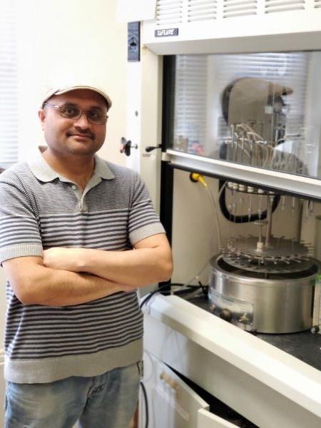
Dr. Poorna Yalagala, Postdoctoral Research Associate at the University of Illinois at Chicago is no stranger to working with Organomation nitrogen blowdown evaporators. He and his colleague have relied on Organomation N-EVAP sample dryers for their research for 15 years. He was first introduced to Organomation N-EVAPs while pursuing his PhD in India at the Central Food Technological Research Institute (CSIR). Today, Dr. Yalagala is using the N-EVAP’s for his research into the potential role of dietary docosahexaenoic acid (DHA) in brain function and prevention of cardiovascular diseases and neurological disorders.
Application: Impact of DHA on Brain Function of Mice
Gas Chromatography-Mass Spectrometry (GC-MS) is a powerful analytical technique used extensively in biochemical research, including studies involving dietary docosahexaenoic acid (DHA) and brain function in mice. Here’s how GC-MS is typically utilized in such research, with an example from the work of Dr. Yalagala and his colleagues:
GC-MS Sample Preparation
1. DHA Preparation for Gavage: Dr. Yalagala and his team prepared DHA samples for gavage by dissolving DHA compounds in chloroform within glass vials. The solvent was then evaporated under nitrogen, and the remaining lipid was dissolved in ethanol before being added to corn oil.
2. Tissue Collection: Brain tissues were collected from mice that had been fed the DHA-enriched diet or a control diet.
3. Lipid Extraction: Lipids were extracted from the brain tissues using organic solvents such as chloroform and methanol, isolating the lipids, including DHA, from other cellular components.
4. Derivatization: To make the fatty acids more volatile and suitable for GC-MS analysis, they were converted into fatty acid methyl esters (FAMEs) through derivatization, typically involving methanol and a catalyst.
GC-MS Analysis
1. Gas Chromatography (GC): The derivatized samples were injected into a Shimadzu GC/MS system (Shimadzu Auto Sampler AOC-20s), where they were vaporized and passed through a chromatographic column. The column separated the compounds based on their volatility and interaction with the column material.
2. Detection and Mass Spectrometry (MS): As the separated compounds exited the GC column, they entered the mass spectrometer. The MS ionized the compounds, usually using electron ionization, and measured the mass-to-charge ratio of the ionized fragments, producing a unique mass spectrum for each compound.
3. Data Analysis: The mass spectra were compared against known spectra in libraries to identify the compounds present. Quantitative analysis was performed to measure the concentration of DHA and other fatty acids in the brain tissue.
Applications in Brain Function Research
1. DHA Quantification: Researchers quantified the levels of DHA in brain tissues to assess how dietary supplementation affected its concentration.
2. Metabolite Profiling: GC-MS allowed for the profiling of various metabolites derived from DHA, providing insights into its metabolic pathways and mechanisms of action in the brain.
3. Comparative Studies: By comparing the lipid profiles of DHA-supplemented and control mice, researchers identified changes in lipid composition that correlated with alterations in brain function.
4. Biomarker Discovery: GC-MS helped identify potential biomarkers related to brain health and DHA metabolism, aiding in understanding how DHA influences brain function.
Advantages of Using GC-MS
- High Sensitivity and Specificity: GC-MS is highly sensitive and specific, allowing for the accurate identification and quantification of DHA and its metabolites even in small brain tissue samples.
- Comprehensive Analysis: It provides a comprehensive analysis of the lipid profile, offering insights into the broader effects of DHA supplementation.
- Reproducibility: The technique is highly reproducible, ensuring consistent results across different samples and experiments.
In summary, GC-MS is an essential tool in researching the role of dietary DHA in brain function. Through meticulous sample preparation and advanced analytical techniques, Dr. Yalagala and his colleagues effectively utilized GC-MS to analyze DHA brain content in mice, providing valuable insights into how dietary DHA impacts brain function.
Conclusion: Organomation’s Instrument Durability Can’t be Beat
Dr. Yalagala looks forward to replacing his lab’s original Organomation Meyer N-EVAP from the 90s. After 30 years, the N-EVAP still works. The N-EVAP design has always been “perfect for [my] needs,” he said. The N-EVAP sample holder can hold any sized test tubes between 10-30 mm in outside diameter. He can’t wait to start using Organomation’s latest 24 position N-EVAP design, with a dry bath, and acid resistant coating, to further his research in the prevention of cardiovascular diseases and neurological disorders.
 |
Last Updated 8/22/24 (Photo: Dr. Poorna Yalagala, Postdoctoral Research Associate at the University of IL at Chicago utilizes Organomation's 24 position N-EVAP for his research on DHA brain function in mice.)
